
Introduction to Platelet Antibody Detection
The detection of platelet antibodies is an essential step in the diagnosis and monitoring of several hematological and transfusion-related disorders. Clinicians require a diagnostic method that is simple, reliable, sensitive, and suitable for routine laboratory applications, similar to the antiglobulin test commonly used for red blood cells. Traditional platelet antibody detection methods such as the platelet agglutination test (PAT) and complement fixation test (CFT) are relatively easy to perform and useful for identifying platelet and HLA antigens. However, these conventional assays fail to detect non-agglutinating and non-complement-binding antibodies, which are frequently involved in clinically important conditions.
This limitation becomes particularly problematic in patients receiving repeated platelet transfusions, individuals with idiopathic thrombocytopenic disorders, and newborns affected by neonatal alloimmune thrombocytopenia. In these cases, clinically significant antibodies may remain undetected, reducing diagnostic accuracy and compromising patient management.
Although methods such as the antiglobulin consumption test, radioactive antiglobulin assays, quantitative fluorescence techniques, and immunofluorescence microphotometry have improved sensitivity, these techniques are often labor-intensive, technically demanding, and unsuitable for large-scale routine diagnostic use. Therefore, there has been a strong need for a simplified yet highly sensitive laboratory method capable of identifying both platelet-specific alloantibodies and autoantibodies.
Development of the Platelet Suspension Immunofluorescence Test (PSIFT)
Researchers observed that nonspecific fluorescence, a major limitation in platelet immunofluorescence assays, could be significantly reduced through fixation of blood cells with paraformaldehyde (PFA). This discovery emerged during studies involving heteroantisera production and granulocyte antibody detection.
When platelets were treated with paraformaldehyde before immunofluorescence analysis, unwanted background fluorescence was almost completely eliminated. This improvement allowed the development of a new diagnostic assay known as the Platelet Suspension Immunofluorescence Test (PSIFT).
The PSIFT method was specifically designed to provide:
- High sensitivity for platelet antibody detection
- Reduced nonspecific fluorescence
- Improved reproducibility
- Compatibility with routine laboratory workflows
- Detection of both IgG and IgM platelet antibodies
The assay became particularly valuable because it successfully detected platelet-reactive antibodies that were missed by traditional agglutination and complement fixation methods.
Materials Used in the PSIFT Method
Serum Samples
Different categories of sera were used during the development and validation of the assay:
- Normal sera from healthy male donors served as negative controls
- Human complement sources were prepared from pooled AB sera
- Platelet-specific antisera were collected from patients with post-transfusion purpura and neonatal thrombocytopenia
- Drug-dependent antibody sera containing quinine- or quinidine-dependent antibodies were included
- Autoimmune platelet antibody sera were also tested
Additional antisera against HLA antigens, ABO blood groups, Rh antigens, and granulocyte-specific antigens were evaluated to assess assay specificity.
Fluorescent Antiglobulin Reagents
Fluorescein isothiocyanate (FITC)-labeled antiglobulin reagents targeting:
- Total immunoglobulins
- IgG
- IgM
- Complement component C3
were used to visualize antibody binding on platelet surfaces.
Paraformaldehyde Fixation Solution
A 4% paraformaldehyde stock solution prepared in phosphate-buffered saline (PBS) was diluted to a working concentration of 1% before platelet fixation. Proper pH adjustment and dark storage conditions were critical for maintaining reagent stability and performance.
Preparation of Paraformaldehyde-Fixed Platelets
Platelets were isolated from EDTA-treated blood samples obtained from healthy donors, preferably blood group O individuals to minimize interference from ABO antibodies.
The preparation process included:
- Centrifugation to obtain platelet-rich plasma
- Washing platelets with EDTA-PBS buffer
- Fixation with 1% paraformaldehyde for 2–5 minutes at room temperature
- Additional washing steps to remove excess fixative
- Resuspension of platelets at suitable concentrations for testing
Although approximately 50% of platelets were recovered after fixation, the resulting suspension produced stable and reproducible immunofluorescence results.
Importantly, paraformaldehyde-fixed platelets retained their antigenic properties and could still bind platelet-specific antibodies efficiently.
Procedure of the Platelet Suspension Immunofluorescence Test
The PSIFT protocol involves several sequential incubation and washing steps:
Step 1: Incubation with Patient Serum
Fixed platelets are mixed with patient serum and incubated at 37°C for 30 minutes, allowing platelet antibodies to bind surface antigens.
Step 2: Washing Procedure
Platelets are washed multiple times with EDTA-PBS buffer to remove unbound serum proteins and reduce nonspecific reactions.
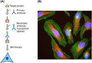
Step 3: Addition of FITC-Labeled Antiglobulin
Fluorescent antiglobulin reagents are added to detect bound antibodies on platelet membranes.
Step 4: Microscopic Examination
After final washing and mounting, fluorescence is examined under a fluorescence microscope. Positive samples show bright ring-like fluorescence surrounding platelets, indicating antibody binding.
For complement studies, an additional incubation step with human complement and FITC-labeled anti-C3 reagent is performed.
Prevention of Nonspecific Fluorescence
One of the major achievements of the PSIFT method was the elimination of nonspecific fluorescence, which had previously limited platelet immunofluorescence assays.
Paraformaldehyde fixation likely reduces nonspecific fluorescence through:
- Modification of membrane protein amino groups
- Increased negative membrane charge
- Reduced nonimmune immunoglobulin binding
- Possible inactivation of Fc receptors on platelet surfaces
This fixation process dramatically improved assay specificity while preserving true antigen-antibody interactions.
Specificity of the PSIFT Assay
The PSIFT demonstrated high specificity for platelet antibodies.
Detection of Platelet-Specific Alloantibodies
The assay successfully detected antibodies against platelet antigens such as:
- Zw(a)
- Zw(b)
- Ko(a)
- Ko(b)
Importantly, non-agglutinating IgG antibodies undetectable by platelet agglutination tests were clearly identified using PSIFT.
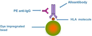
HLA Antibody Detection
The assay also detected HLA antibodies bound to platelet surfaces. Some sera produced additional positive reactions due to contaminating HLA antibodies not identified by conventional lymphocytotoxicity tests.
ABO and Rh Antibody Testing
- Anti-A and anti-B sera reacted with platelets carrying corresponding ABO antigens
- Anti-D sera showed no platelet reactivity, confirming assay specificity
Granulocyte Antibody Testing
Granulocyte-specific antisera without HLA contamination showed negative PSIFT reactions, demonstrating minimal cross-reactivity.
Sensitivity of the Platelet Immunofluorescence Assay
The PSIFT showed superior sensitivity compared with traditional platelet agglutination and complement fixation assays.
Detection of IgG Platelet Antibodies
IgG antibodies such as anti-Zw(a), which fail to induce visible platelet agglutination, produced strong fluorescence signals in the PSIFT assay. This confirmed the ability of the method to detect incomplete or non-agglutinating antibodies.
Drug-Dependent Antibody Detection
Quinine- and quinidine-dependent platelet antibodies were detected at significantly higher titers using PSIFT compared with complement fixation methods.
HLA Antibody Sensitivity
For highly selected HLA antisera, the sensitivity of PSIFT was comparable to:
- Lymphocytotoxicity tests
- Lymphocyte immunofluorescence tests
- Granulocyte immunofluorescence assays
Reproducibility and Reliability
Repeated testing demonstrated excellent reproducibility of the PSIFT assay.
- No false-positive reactions were observed
- No false-negative reactions occurred
- Positive and negative control sera produced consistent results over hundreds of experiments
This high reliability supports the suitability of PSIFT for routine clinical laboratory use.
Quantification of Platelet Immunofluorescence
Researchers also evaluated platelet fluorescence quantitatively using continuous-flow microfluorometry.
The Biophysics Cytofluorograph measured:
- Fluorescence intensity per platelet
- Distribution of fluorescence across thousands of cells
- Relative fluorescence intensity (RFI)
This quantitative approach demonstrated clear differentiation between antigen-positive and antigen-negative platelets, suggesting potential applications in:
- Platelet antigen quantification
- Antibody titration
- Automated diagnostic analysis
Clinical Importance of the PSIFT Method
The Platelet Suspension Immunofluorescence Test offers several major clinical advantages:
Detection of Clinically Relevant IgG Antibodies
The method efficiently identifies non-agglutinating IgG platelet antibodies involved in:
- Neonatal alloimmune thrombocytopenia
- Autoimmune thrombocytopenia
- Platelet transfusion refractoriness
Improved Diagnostic Accuracy
By detecting antibodies missed by traditional assays, PSIFT enhances diagnostic sensitivity and supports better patient management.
Routine Laboratory Applicability
Unlike radioactive or highly specialized methods, PSIFT is relatively simple, reproducible, and practical for large-scale clinical laboratory implementation.
Conclusion
The development of the Platelet Suspension Immunofluorescence Test represents a major advancement in platelet immunology and transfusion medicine. By combining paraformaldehyde-fixed platelets with fluorescence-based antibody detection, researchers created a highly sensitive and reproducible assay capable of identifying both IgG and IgM platelet antibodies.
The method successfully overcomes the limitations of conventional platelet agglutination and complement fixation techniques, particularly in the detection of non-agglutinating antibodies associated with serious hematological disorders.
Due to its simplicity, specificity, sensitivity, and compatibility with routine laboratory workflows, PSIFT has become a valuable diagnostic tool for investigating platelet alloimmunization, autoimmune thrombocytopenia, neonatal thrombocytopenia, and platelet transfusion complications.